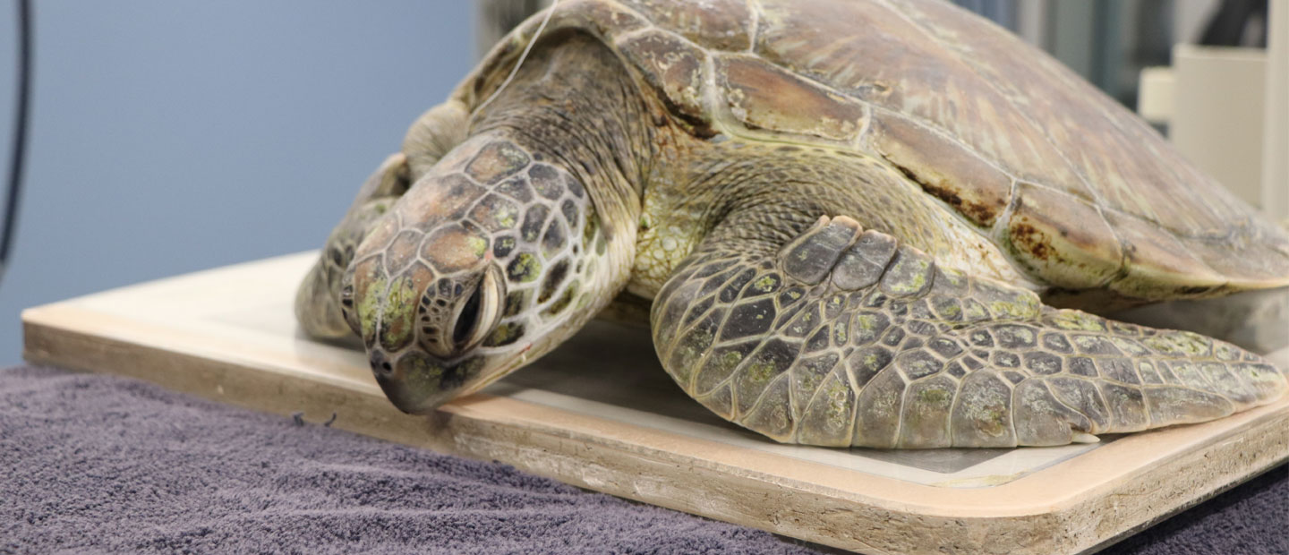
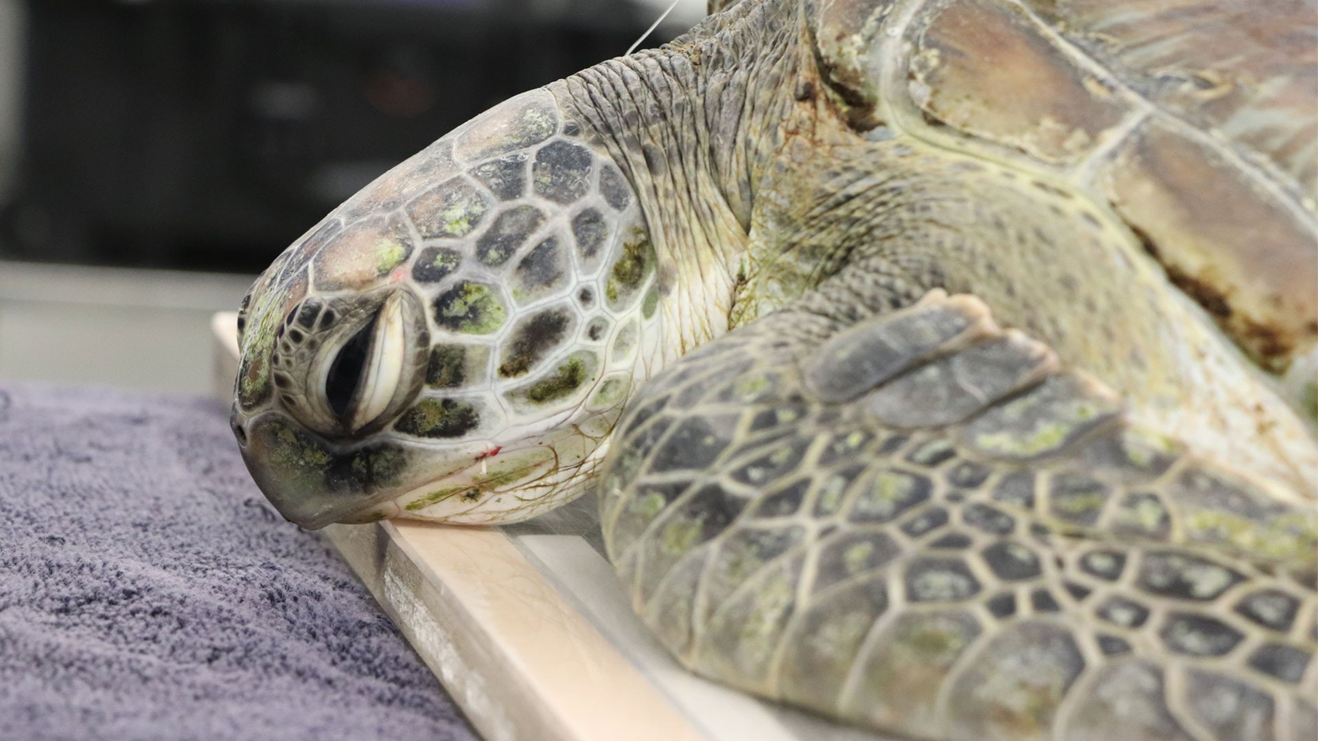
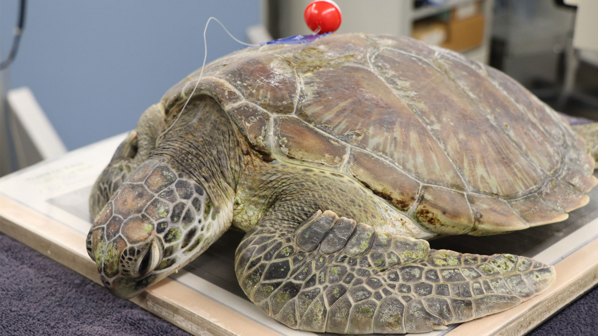
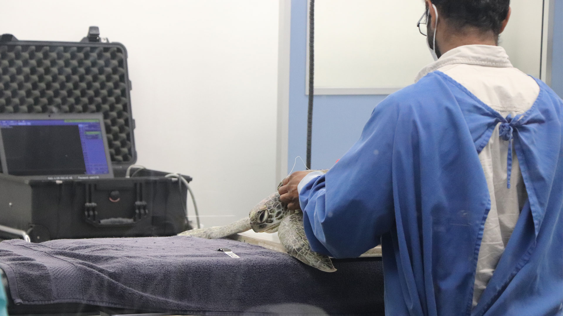
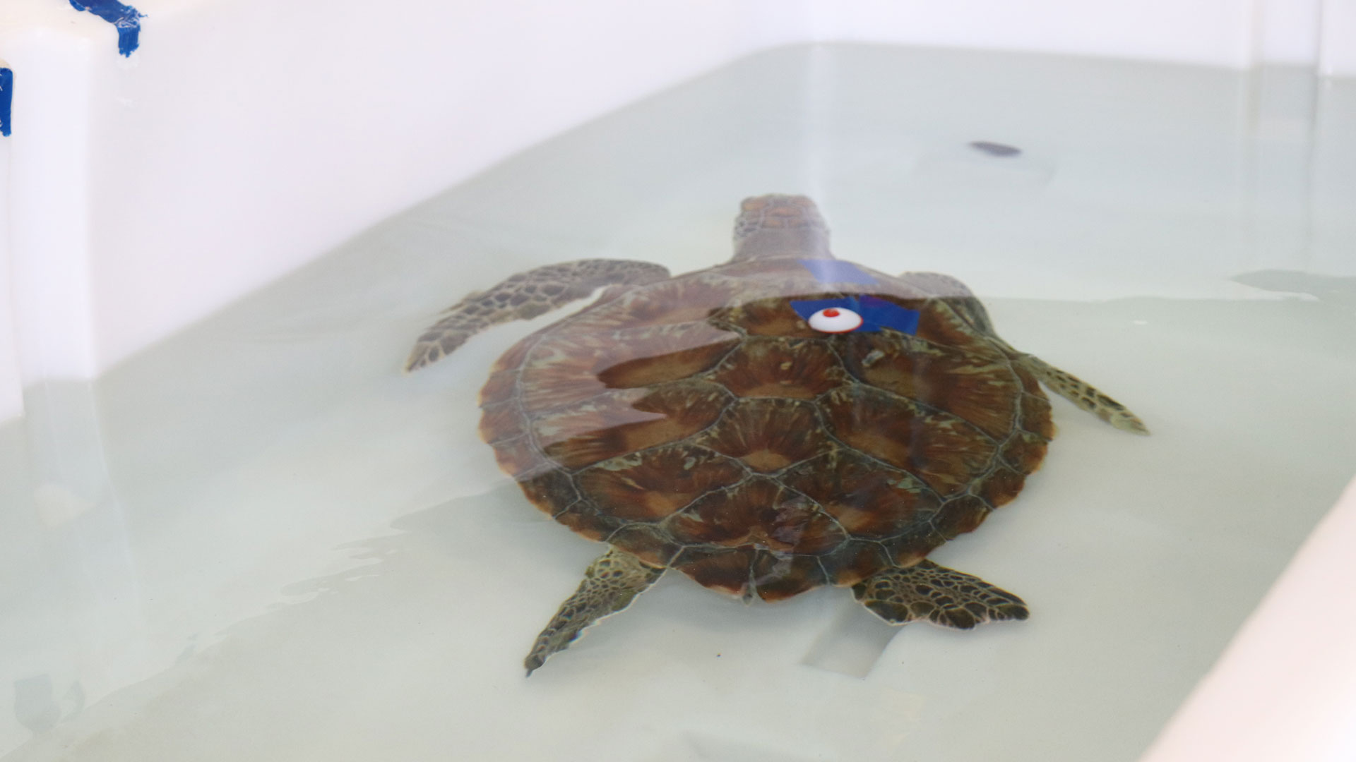
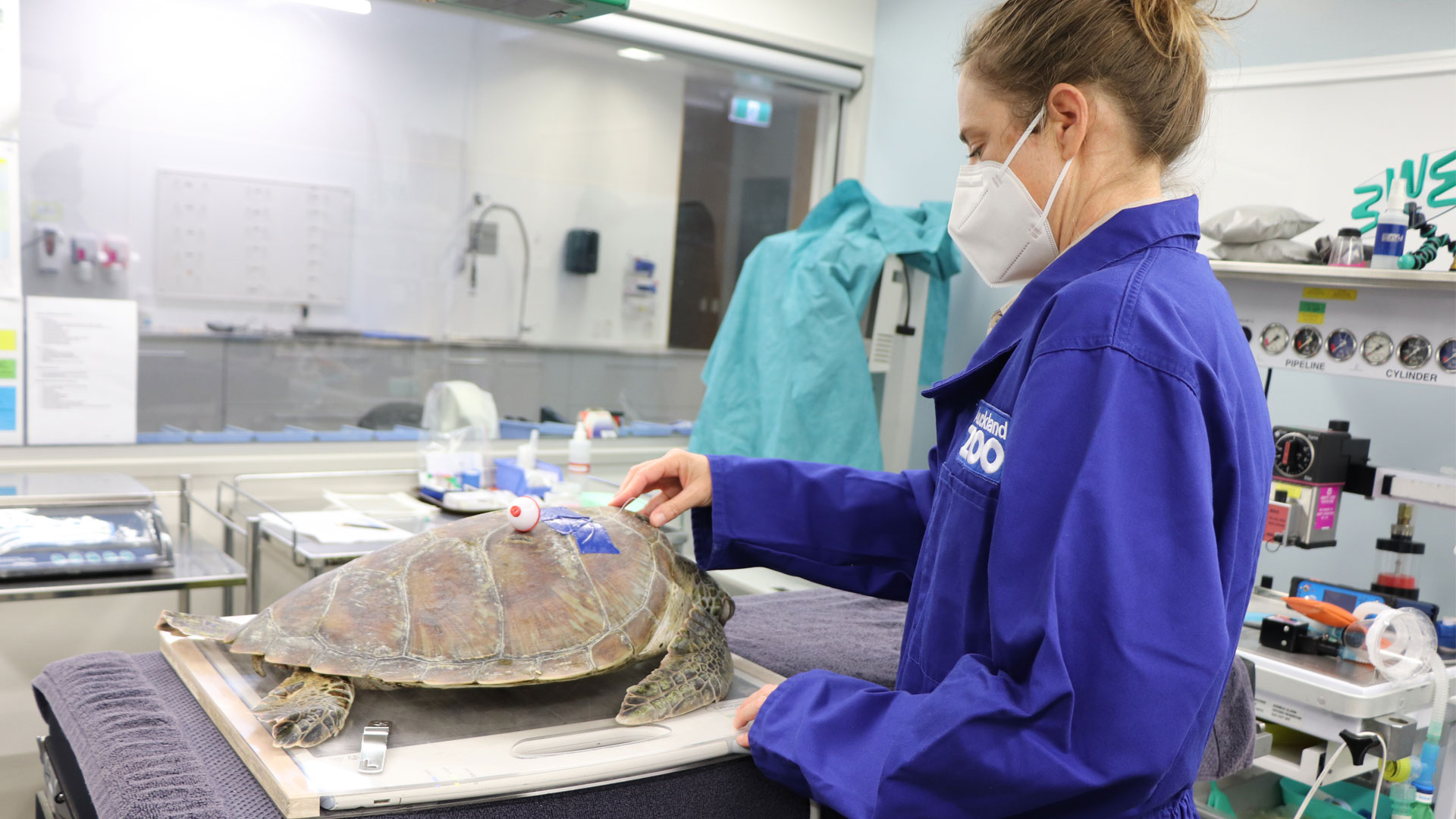
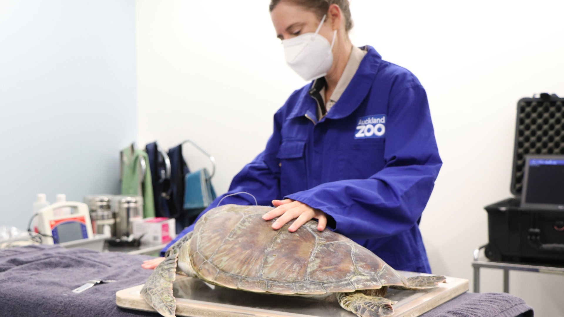
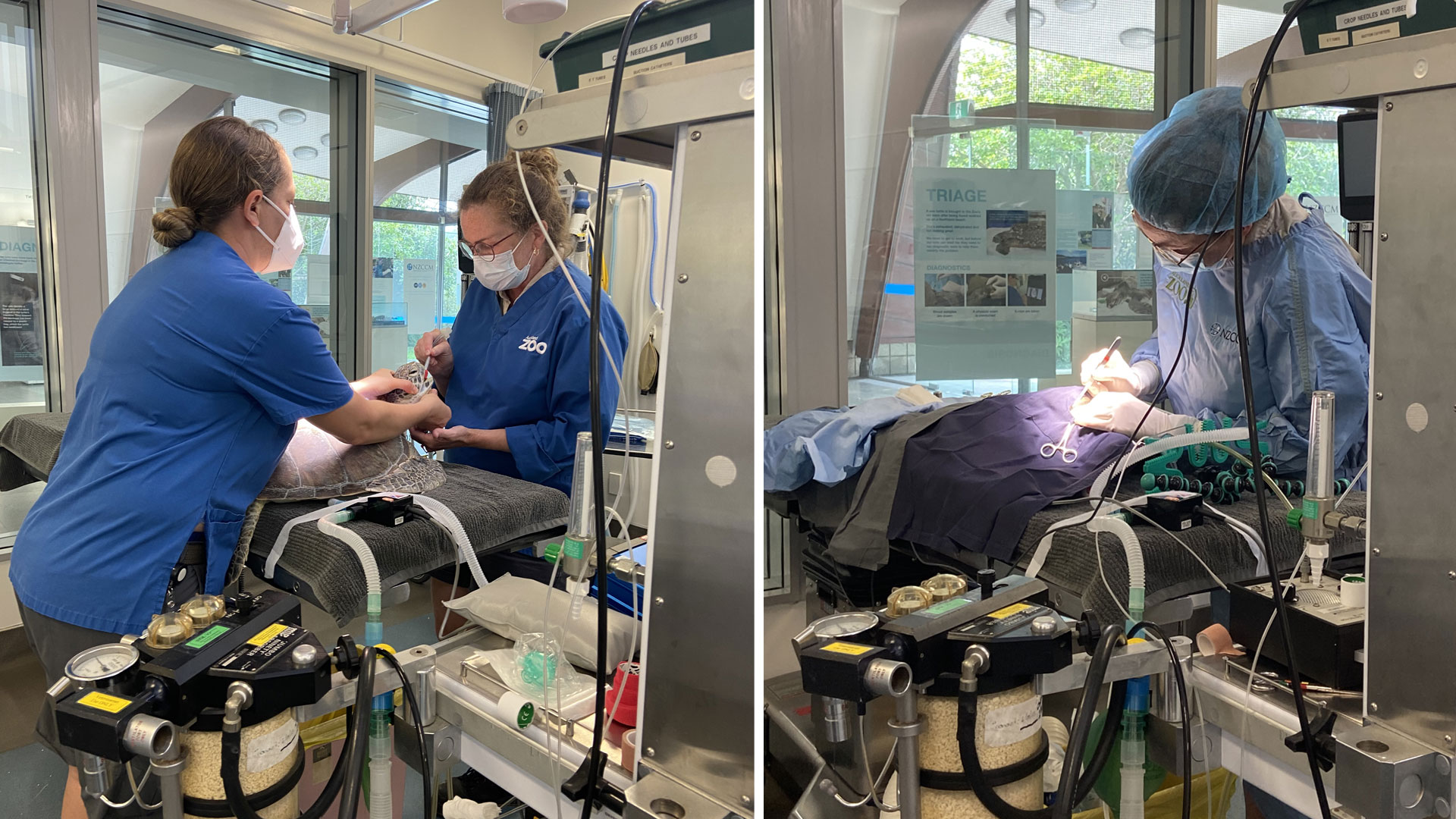
“The fishermen did everything right in this situation – they contacted DOC immediately and followed advice not to cut the fishing line short. Instead, they left about 30cm of the line trailing out of the turtle’s beak and attached it to the carapace (shell) so it wouldn’t be swallowed. Often the fishing line does more damage to the digestive tract than the hook itself and it can’t be seen on X-ray, so this gave the turtle an even better chance of survival”, says vet nurse Celine.
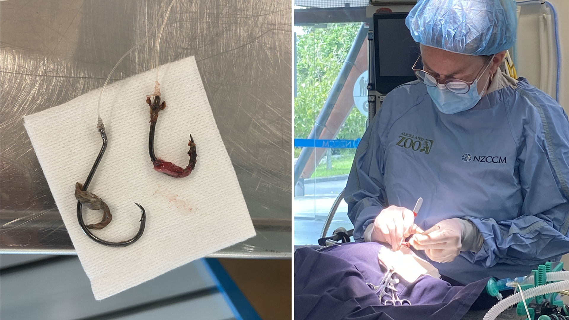
When the sub-adult turtle first arrived at the zoo, it was assessed by our team which included taking blood samples to get an overall state of health, and preliminary x-rays to assess the placement of the hook. This revealed a surprise – as well as the known hook, there was also a second, older hook in the turtle’s oesophagus that must have been consumed previously.
After stabilising the turtle, our vet team anaesthetised their patient and then assessed the damage using an endoscope which showed both hooks had pierced the wall of the oesophagus. To plan for surgery, comprehensive 3D imaging of the exact location of both hooks was required, so the sea turtle was carefully transported in the zoo ambulance to Veterinary Specialists Auckland to have a CT scan (a computerized tomography scan uses Xrays to image the body in 3D). These images were interpreted by Dr. Dennison-Gibby (a specialist veterinary radiologist) who could see that the older hook had a thick layer of abnormal tissue around it and the point of one hook was within a few mm of the heart and lungs. Despite this, surgical removal was thought to be tricky but possible.


